Fdp Tendon Anatomy : Flexor Tendon Injuries Radsource : Flexor tendon pulley has been very early noticed and described.
Get link
Facebook
X
Pinterest
Email
Other Apps
Fdp Tendon Anatomy : Flexor Tendon Injuries Radsource : Flexor tendon pulley has been very early noticed and described.. Flexor tendon pulley has been very early noticed and described. Fd p is at the p oint of the finger). Flexor digitorum profundus muscle is a powerful flexor of the fingers. Together the flexor pollicis longus, pronator quadratus, and flexor digitorum profundus form the deep layer of ventral forearm muscles. Edc tendons straighten the index, middle, ring and small fingers.
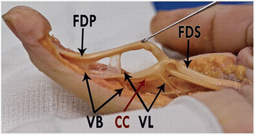
The fds tendon divides into two slips that wrap around the fdp to insert into the sides of the middle phalanx. When a muscle contracts, the tendon pulls on the bone causing the joint to move. Note the joining of the two slips (arrows) before their final individual insertions. The flexor digitorum profundus is a muscle in the forearm of humans that flexes the fingers (also known as digits). This region includes the distal part of the fdp tendon.
Suture Fixation For Distal Phalanx Base Palmar Avulsion from resources.aofoundation.org Flexor digitorum profundus muscle is a powerful flexor of the fingers. Micro and macro anatomy, biology of tendon healing This contains the superficial and deep flexor tendons, flexor pollicis longus tendon and the median nerve. Terminology usually accepted recognizes 6 arcifom pulleys (a0 to a5) and 3 cruciform pulleys (c1 to c3). (b) photograph of a dissected anatomic specimen shows a superficial volar view of the chiasm of the fds tendon. The reconstruction of chronic flexor tendon injuries remains one of the more challenging injuries facing the hand and upper extremity surgeon. It is considered an extrinsic hand muscle because it acts on the hand while its muscle belly is located in the forearm. Together the flexor pollicis longus, pronator quadratus, and flexor digitorum profundus form the deep layer of ventral forearm muscles.
Fd p is at the p oint of the finger).
Terminology usually accepted recognizes 6 arcifom pulleys (a0 to a5) and 3 cruciform pulleys (c1 to c3). Note the joining of the two slips (arrows) before their final individual insertions. The tendons of the fdp and fds lie deep in the palm of the hand, beneath the superficial palmar arch and the superficial branches of the median and ulnar nerve. Flexor digitorum superficialis (fds) and flexor digitorum profundus (fdp). The largest compartment is the carpal tunnel. Anatomy of the flexor tendons. Damage to the foot and ankle tendons are a common cause of foot pain, typically caused by overuse, overstretching or an injury. Flexor digitorum superficial splits into radial and ulnar slips prior to insertion on middle phalanx. Zone 1 is distal to the flexor digitorum superficialis insertion (fds) and contains only the flexor digitorum profundus (fdp), zone 2 contains both the. This region includes the distal part of the fdp tendon. As with tendons elsewhere, the extensor tendons are best assessed in the axial plane. The fds tendons are the most palmar, and the fds tendons to the long and ring fingers are most superficial. Flexor digitorum profundus muscle is a powerful flexor of the fingers.
The fds tendon divides into two slips that wrap around the fdp to insert into the sides of the middle phalanx. Related posts of thumb flexor tendon anatomy anatomy shoulder bones diagrams. The largest compartment is the carpal tunnel. Anatomy shoulder bones diagrams 11 photos of the anatomy shoulder bones diagrams anatomy ankle bones, anatomy elbow bones, anatomy hip bones, anatomy knee bones, anatomy scapula, anatomy shoulder muscles, anatomy shoulder pain, parts of shoulder joint, hand, anatomy ankle bones, anatomy elbow bones, anatomy hip. Muscles flexor digitorum profundus (fdp) functions as a flexor of the dip joint.
Zone Ii Flexor Tendon Repair Musculoskeletal Key from musculoskeletalkey.com This region includes the distal part of the fdp tendon. The vascular anatomy of the flexor digitorum profundus (fdp) tendon insertion is described by using a vascular injection and modified spalteholtz tissue clearing protocol in 36 human cadaver digits. As with tendons elsewhere, the extensor tendons are best assessed in the axial plane. Fd p is at the p oint of the finger). Flexor digitorum superficialis (fds) and flexor digitorum profundus (fdp). Related posts of thumb flexor tendon anatomy anatomy shoulder bones diagrams. The reconstruction of chronic flexor tendon injuries remains one of the more challenging injuries facing the hand and upper extremity surgeon. Which of the following techniques uses the least amount of suture necessary to prevent gap.
Flexor digitorum superficialis is the largest muscle of the anterior compartment of the forearm.
A common muscle belly is shared by. The largest compartment is the carpal tunnel. The flexion loss of the dip joint is seen due to fdp tendon injury. Related posts of thumb flexor tendon anatomy anatomy shoulder bones diagrams. Edc tendons straighten the index, middle, ring and small fingers. Anatomy and physiology of this flexor tendon gliding and reflection system at the level of the digital sheet are exposed. The reconstruction of chronic flexor tendon injuries remains one of the more challenging injuries facing the hand and upper extremity surgeon. Ebraheim's educational animated video describes the anatomy of the flexor digitorum profundus muscle.the flexor digitorum profondus is a muscle in the fo. Note the joining of the two slips (arrows) before their final individual insertions. The flexor tendons of the digits enter the carpal tunnel in a generally consistent anatomic relationship. The lumbricals of the hand arise from the radial side of its tendons. The anatomy of the flexor surface of the wrist is defined principally by the flexor retinaculum. Muscles flexor digitorum profundus (fdp) functions as a flexor of the dip joint.
The reconstruction of chronic flexor tendon injuries remains one of the more challenging injuries facing the hand and upper extremity surgeon. The flexor tendons of the digits enter the carpal tunnel in a generally consistent anatomic relationship. This contains the superficial and deep flexor tendons, flexor pollicis longus tendon and the median nerve. This region includes the distal part of the fdp tendon. The fdp tendon is not present.
Flexor Tendon Injuries Of The Hand Ppt Video Online Download from slideplayer.com Each finger is connected to the muscles by two tendons, the flexor profundus (fp) and the flexor superficialis (fs). The flexor mechanism of the index, middle, ring, and small fingers consists of the flexor digitorum profundus (fdp) and the flexor digitorum superficialis (fds) tendons. Anatomy and physiology of this flexor tendon gliding and reflection system at the level of the digital sheet are exposed. The tendons on the flexor side of the wrist are also arranged in compartments. Which of the following techniques uses the least amount of suture necessary to prevent gap. Tendon injuries are categorized by location: Ebraheim's educational animated video describes the anatomy of the flexor digitorum profundus muscle.the flexor digitorum profondus is a muscle in the fo. The lumbricals of the hand arise from the radial side of its tendons.
The apl tendon runs on the.
Anatomy and physiology of this flexor tendon gliding and reflection system at the level of the digital sheet are exposed. The fdp and fpl tendons are found in the deepest level of the carpal tunnel. The fdp tendon is not present. Flexor digitorum profundus muscle is a powerful flexor of the fingers. The flexor mechanism of the index, middle, ring, and small fingers consists of the flexor digitorum profundus (fdp) and the flexor digitorum superficialis (fds) tendons. Zone 1 is distal to the flexor digitorum superficialis insertion (fds) and contains only the flexor digitorum profundus (fdp), zone 2 contains both the. The largest compartment is the carpal tunnel. Note the joining of the two slips (arrows) before their final individual insertions. The reconstruction of chronic flexor tendon injuries remains one of the more challenging injuries facing the hand and upper extremity surgeon. Related posts of thumb flexor tendon anatomy anatomy shoulder bones diagrams. Flexor tendon pulley has been very early noticed and described. The fp connects to and controls the motion of the distal phalange (the most distant bone in each finger) and the fs connects to and controls the motion of the middle phalange (the next bone down). Flexor digitorum superficial splits into radial and ulnar slips prior to insertion on middle phalanx.
There, the tendons are surrounded by several layers of loose connective tissue fdp tendon. Each finger is connected to the muscles by two tendons, the flexor profundus (fp) and the flexor superficialis (fs).

Comments
Post a Comment